Anatomy Models Specification
- Material
- Paper and Plastic
- Color
- Olive Green and Pink
Anatomy Models Trade Information
- Minimum Order Quantity
- 1 Piece
- Payment Terms
- Cash in Advance (CID), Cash Advance (CA)
- Supply Ability
- 15 Pieces Per Day
- Main Export Market(s)
- Australia, North America, South America, Eastern Europe, Middle East, Western Europe, Africa, Central America, Asia
- Main Domestic Market
- All India
About Anatomy Models
29 Human EyeVertical Section Greatly Enlarged
Showing Muscle Optic Nerves Crystaline Lens Iris
Cornea etc
30 Human Eye ball100 times enlarged
Detatchable
31 Visual Central Nervous System Pathways
Superior View
32 EarLarge SizeDissectable in 4 Parts
33 Structure within the inner ear including the
cochlea Vastibular Apparatus
34 EarSagittal SectionOn board
External Middle Inner Ear
35 LarynxAnterior View Posterior View
Side View Cut away Side View
Sagittal Section 5 Models
36 Functional Model of Larynx
37 LarynxDeep Side View
38 The PharynxPosterior View
39 Pharynx Sagittal Section
40 TonsilsPharyngeal Palatine
Lingual Tonsils
41 Teeth Lower jaw with structure shown
42 The Structure of tooth
43 The Cavity in tooth
44 The TongueDorsal Surface
45 Pituitary GlandHypothalamus
46 Thyroid Parathyroid Glands
47 Sagittal Section through Nasal Cavity
and Pharynx Viewed from Medial Side
48 LungsOneside sectioned with
Respiratory Tract Bronchial
Tubes Arteries Veins
49 Pulmonary circulation
50 The Respiratory System
51 LiverEnlarged showing Gall Bladder
52 Liver with Gall Bladder Pancreas On Stand
53 Blood Supply of the Liver
54 Duct System with Gall Stones in common sites
55 The Hepatic Portal System
56 Endocrine System
57 Pancreas Enlarged
58 Structure of the Pancreas
59 StomachEnlargedwith duodenum
sectioned showing details
60 An Anterior view of Abdominal aorta
Its principles branches
61 Spleennormal size with details
62 Gall Bladder Pancreas Duodenum
63 Blood Supply of the Intestine
64 Rectum Anal Canal
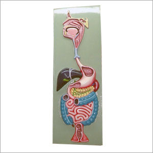
65 Large Intestine
66 Small Intestine
67 The Digestive System
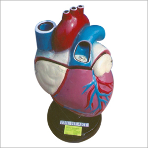
68 HeartEnlargedSeparable in 4 Parts
69 Fat deposition in the arteries
70 Death of an Artery
71 Artery sections with Blockage
Plaque builtup on artery walls
72 Principal Arteries of the body
73 Principal Veins of the body
74 Veins that drain the head Neck


 Send Inquiry
 Send Inquiry
 Send SMS